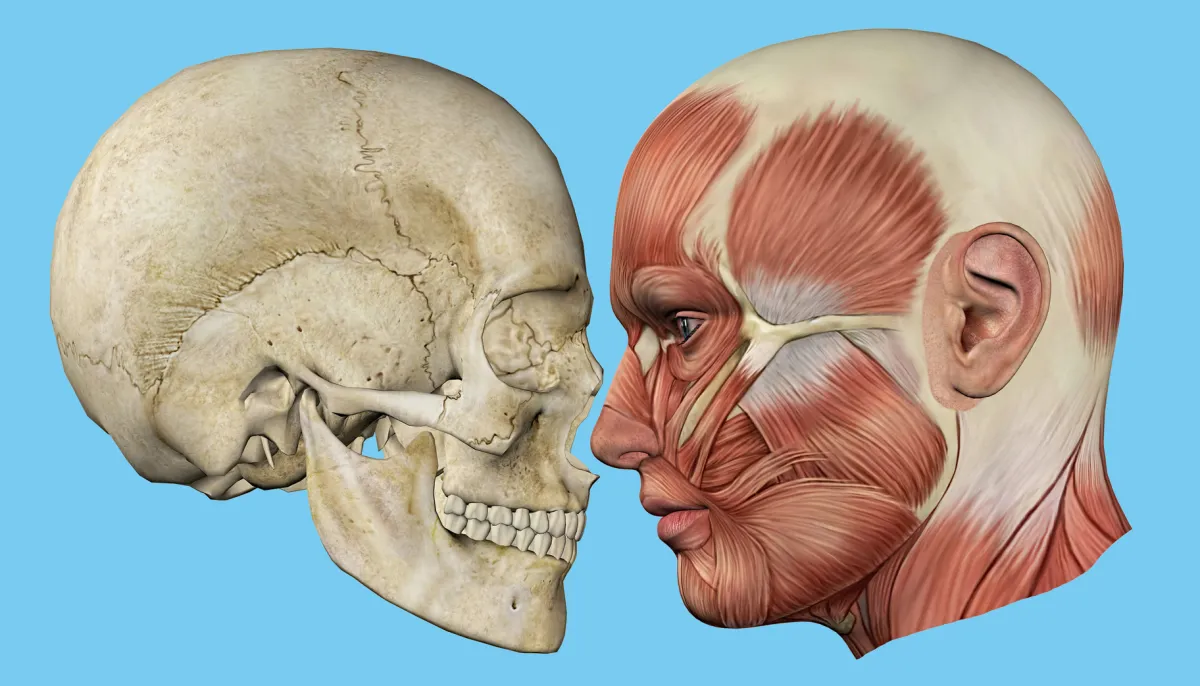
If you’re looking for a gorgeous way to decorate your walls, consider hanging a Skull Side Profile wall art. These wall decor items are available in various materials, from framed to unframed, and in various sizes. In addition, you can find a variety of gallery-quality art prints of this popular design.
Various anatomical features of the skull
The skull has anatomic features, including foramina, that allow nerves and blood vessels to pass through it. These openings are called fontanelles, allowing the brain to grow and develop. The anterior and posterior fontanelles close at about the same age, while the middle and lateral foramina open at different ages. Each of these openings has a different shape and size.
The human skull consists of twenty-two bones joined by cranial sutures. The base of the skull is sub-categorized into the anterior, middle, and posterior cranial fossae and consists of the frontal, ethmoid, sphenoid, occipital, and temporal bones. The bones of the face are also found in the skull and are collectively known as the facial skeleton. The palatine and lambdoid bones are part of the frontal and occipital bones.
Bony elements of the skull
The bones that comprise the human skull are known as cranial bones. These bones are found on both the skull’s frontal and back. Each of these bones has specific roles within the skull, such as supporting and protecting the brain and central nervous system. They also house various sinuses, as well as various neurovascular structures.
The skull is supplied with blood from the common carotid and vertebral arteries. The external carotid artery is the most important blood supply for the skull and runs up the side of the neck. It has eight major branches, including the maxillary artery and the middle meningeal artery. When these branches align with one another, the face appears normal.
Bony regions of the skull
A network of sutures attaches to the bones of the skull. The coronal suture runs side-to-side across the skull and joins the frontal bone with the parietal bones. The sagittal suture extends posteriorly from the coronal suture and runs along the midline of the top of the skull. This suture joins the right and left parietal bones. The lambdoid suture extends laterally away from this suture.
The outermost portion of the skull consists of five large bones called the temporal, occipital, and frontal bones. The frontal bone forms the front of the skull, while the temporal bone forms the back of the skull. The occipital bone forms the sides of the skull and contains some of the neck muscles. The nuchal lines are located adjacent to these two bones.
Number of bones in the skull
The human skull contains 29 bones and is divided into four main categories:
- The cranial bones (frontal, occipital, sphenoid, and temporal)
- Facial bones (maxilla and mandible)
- Associated bones (hyoid, incus, and lacrimal)
Six bones in the ear are associated with the bones of the head and face, which form the eardrums.
During the development of a human being, the skull is made up of eight cranial bones, including the skull bone. In addition, there are 14 bones in the face, which make up the cheeks, nose, mouth, and jaw. The bones of the nose are relatively small; there are two nasal bones, or conchae, on each side. In addition, there is a third bone, the vomer, which is part of the nasal septum and connects to the cranial bones, the ethmoid bone, and the hyoid bone. The lacrimal bones attached to the lacrimal glands are another set of bones in the face.
Variation in skull profile
Variation in skull profile is a significant trait of some species. The male outer cranial vault exhibits dramatic changes, especially in the temporal, inferior parietal, and posterior parietal regions. In contrast, the female outer cranial vault shows less variation. However, the male and female outer cranial vaults show significant compression at the frontal and midline.
The present study aimed to develop an algorithm to identify a grid of homologous pseudo-landmarks on the skull. The analysis was performed on adults’ cranial base and vault using a 3D geometric morphometric method. These data will access an understanding of the adult skull and the role of different anatomical regions.


